
 TR
TR ES
ES FR
FR AR
AR RU
RULung Nodule Center
In the medical literature, lung nodules—referred to as “pulmonary nodules”—are small clusters of cells found within lung tissue. These structures are usually detected incidentally during chest X-rays or computed tomography (CT) scans performed for other health concerns, and they represent small tissue masses in the lungs. They may occur in a single lung or in both lungs simultaneously. The vast majority of these nodules are benign. However, lesions larger than 25 mm carry a higher risk of being malignant (cancerous) compared to smaller ones.
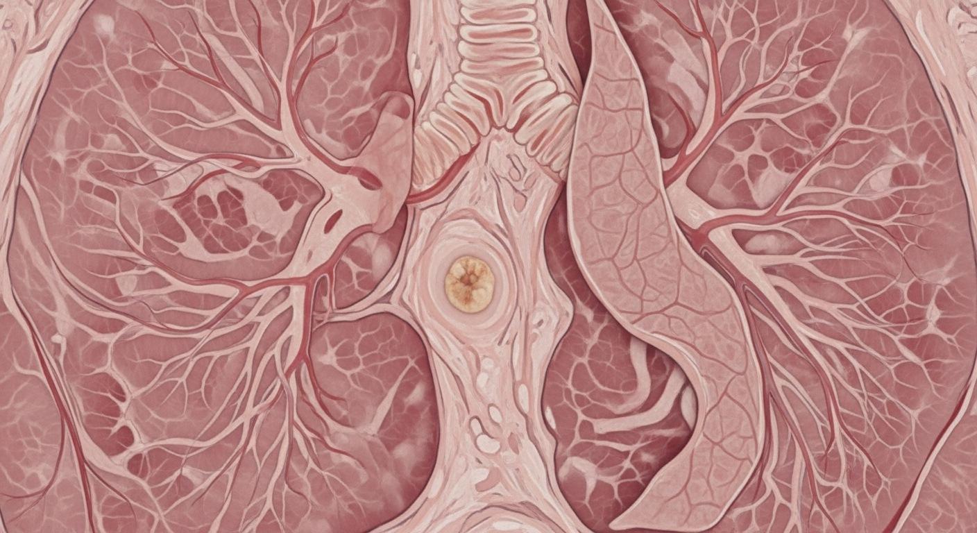
What Is a Lung Nodule?
A lung nodule is an abnormal tissue growth detected in the lung parenchyma, measuring between 1 and 30 millimeters. Lesions larger than 3 centimeters are defined as “masses.” The main characteristic of these structures is that they are firmer and denser than the surrounding normal tissue. Nodules can be either benign or malignant. Most of the time, they do not cause any symptoms and are therefore discovered incidentally. A nodule may be solitary or multiple in number. If the lesion is larger than 6 millimeters and shows a tendency to harden, further evaluations such as radiological follow-up, surgical biopsy, or liquid biopsy through blood tests may be required, considering the risk of cancer. In suspicious cases, surgical removal of the nodule can be life-saving.
Causes of Nodule Formation
During the diagnostic process, the patient’s medical history is of great importance. Bacterial, fungal, or parasitic infections can lead to such changes in the lungs. Tuberculosis (TB), which is relatively common in our country, frequently causes nodules and tissue damage. A person’s history of tobacco use is one of the most critical factors. Malignant lesions are known to be directly associated with smoking. Age and smoking habits are the two most important parameters guiding the physician’s decision-making process. In non-smokers under the age of 35, the probability of cancer is below 1%; whereas in smokers over the age of 55, nodules larger than 15 mm with irregular borders and a firm structure carry an approximately 50% risk of malignancy. In addition, a family history of lung cancer, the presence of emphysema, and the radiological characteristics of the nodule are also taken into consideration.
Classification and Follow-up Lung nodules are radiologically examined in three groups:
- Solid: Completely dense tissue.
- Ground-glass: Low-density, more translucent-appearing areas.
- Part-solid: Structures that contain both solid and ground-glass components.
In accordance with international protocols, the follow-up or treatment approach is determined based on the structure of the nodule. In low-risk cases, periodic monitoring alone may be sufficient, whereas follow-up of ground-glass nodules may extend up to 5 years. This process should be managed under specialist supervision using advanced imaging technologies, without causing unnecessary anxiety for the patient.
Diagnostic Methods
Methods used for the detection and evaluation of nodules include chest X-ray, CT (Computed Tomography), PET/CT, needle biopsy, liquid biopsy, and bronchoscopy, which allows visualization of the airways with a camera. For small lesions under 10 mm, CT is the most suitable follow-up tool. Expert committees compare the patient’s past and current imaging results to determine a personalized management plan.
Liquid Biopsy Technology
Liquid biopsy is an advanced test that detects tumor cells or DNA/RNA fragments circulating in the bloodstream using only 10 ml of blood drawn from the arm. This method does not require a surgical incision and can identify cancerous formations at very early stages. By mapping the genetic profile of the tumor, a precise treatment plan can be established.
Treatment Approaches
Not every detected nodule requires surgical intervention. The patient’s age, the diameter of the nodule (lesions under 6 mm are generally considered safe), its shape, and growth rate are all evaluated. If the risk is deemed high, early diagnosis and removal of the nodule can significantly increase the success of treatment.
Multidisciplinary Approach
Since the lungs are delicate organs with an extensive blood supply, a potential cancer can spread rapidly. Therefore, early diagnosis is critical. At our center, a multidisciplinary team (council) consisting of specialists in Pulmonology, Thoracic Surgery, Radiology, Medical Genetics, and Radiation Oncology evaluates each case from all perspectives and initiates the most appropriate treatment process.
Unit Doctors

Prof. Dr. Mustafa YAMAN